
Written by Sohyun KIM
Research Scientist in AIRS Medical
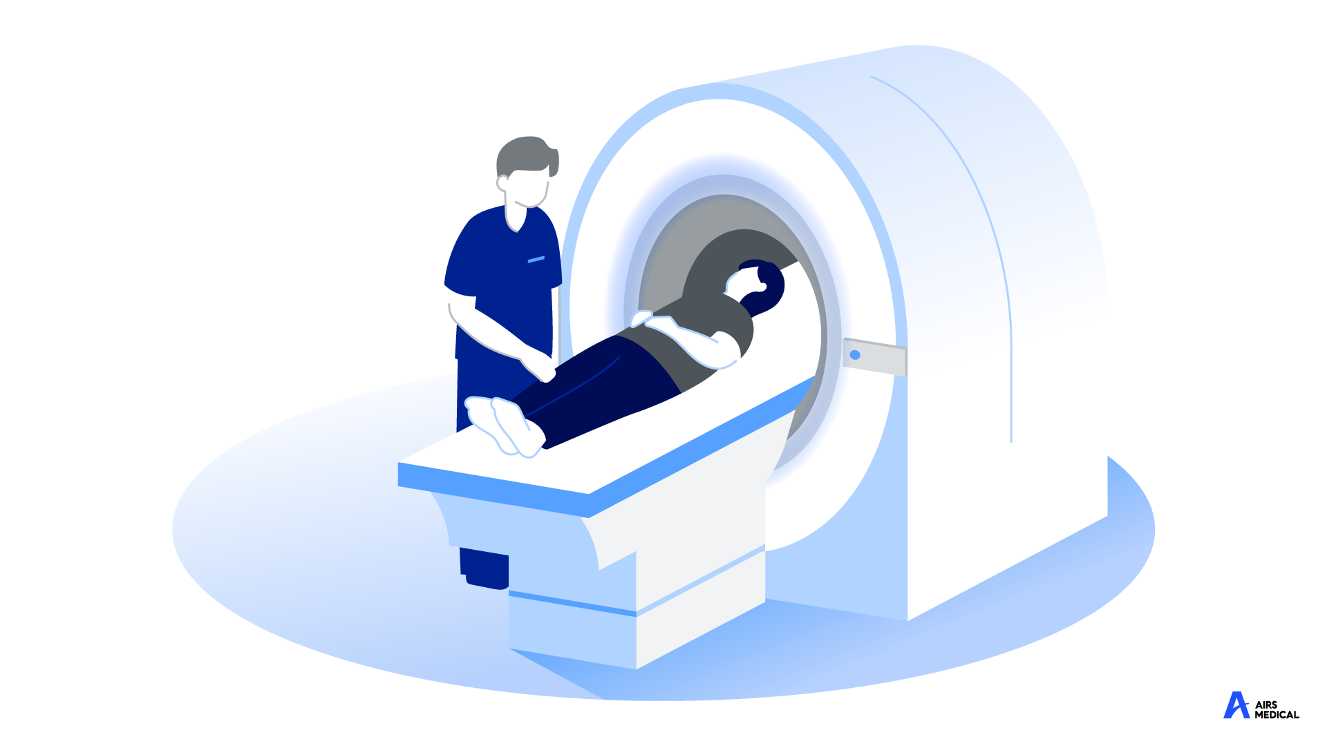
Magnetic Resonance Imaging(MRI), is well-known for imaging that does not use radiation, unlike X-ray or CT scans. MRI can obtain excellent contrast and resolution images of human tissues, so it is a test widely used for diagnosing diseases, determining therapeutic effects, and follow-up.
If you’ve done an MRI scan, you’ve probably been lying on the device thinking, “When will it be over?” X-ray is like taking an ID picture, an ultrasound is done in five minutes, asking the doctor questions, and MRI is usually guided to lie alone in a large device in the examination room without moving for about 30 minutes. I’m already nervous just by being examined at a medical institution, but my whole body is on edge throughout the filming because I’m not moving in a noisy device.
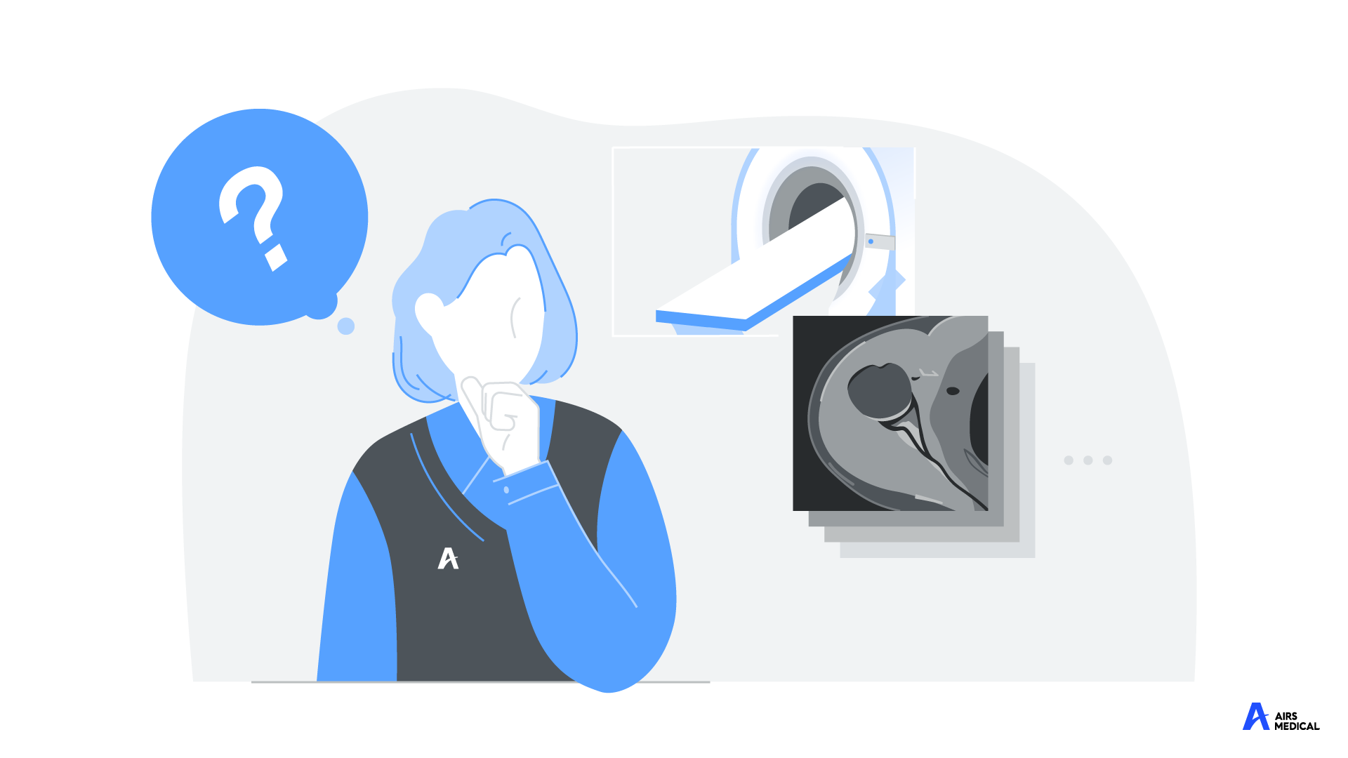
There are several reasons why MRI takes relatively longer than other imaging tests, and one of the most common reasons is that you don’t get a single image during a real MRI. Still, you get different kinds of images from different angles. In the 30-40 minutes or so that we’re being tested, at least three or four different images are being taken of the test site. There is only one area to be examined, and some may wonder why multiple videos are taken. Let’s learn a little more about our bodies and the characteristics of MRI to see why MRI takes a long?
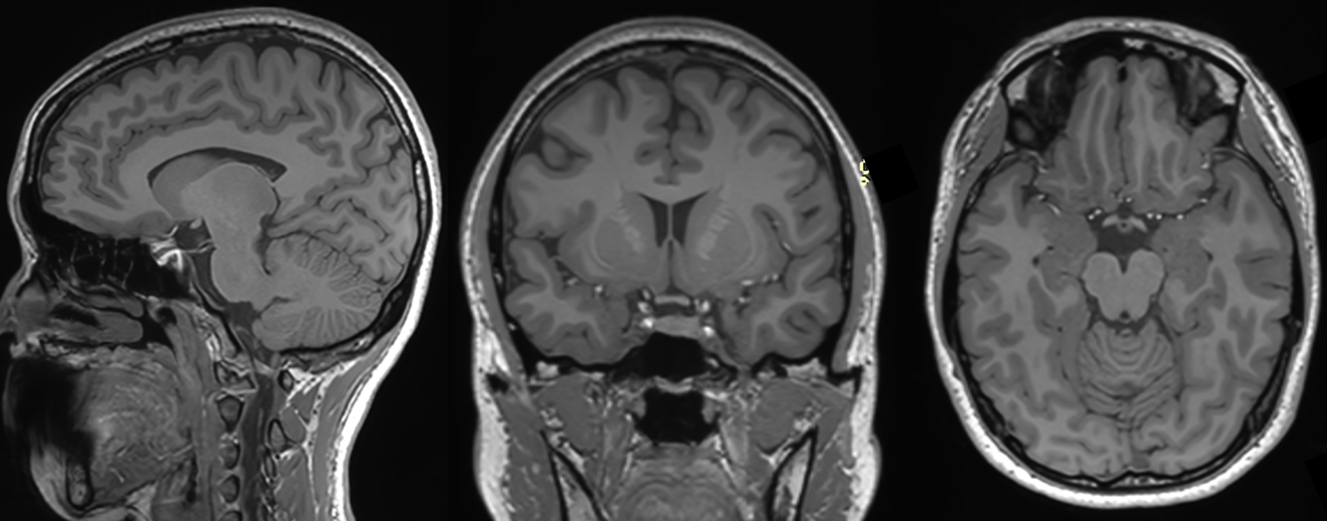
MRI even records three-dimensional spatial information about human structures in receiving and imaging human signals. Shall we look down for a moment? We don’t usually think about it, but our bodies are three-dimensional structures. Next to the thumb is the index finger; if you press the back of your hand, it goes in smoothly and comes out. In the process of receiving signals from the human body, the MRI device records the position of the human structure not only up, down, left, and right, but also the three-dimensional position of the front and back. If you take a picture of one hand on an MRI machine, not only can you see the index finger next to the thumb, but you can also capture the middle of the thickness of the hand, from the back of the hand to the side of the palm. Therefore, when you check the MRI results with your doctor, you can listen to the explanation while moving slowly without stopping the test image in one sheet, like an X-ray image.
Another reason for the increase in scan time is that our body is a three-dimensional structure, and in many cases, MRI images are characterized by only one side at a time. If you have pain in your knee and you want to do an MRI, you’ll have to look at the pain from the side and from the top to find the cause of the pain, but because you get only one side of the image with one MRI, you have to do multiple additional scans to see the same side of the area. The shooting time will increase here.
As discussed earlier, it is not just the concept of the direction or space of the image. Still, there is also a reason why the shooting time increases as various types of images are taken depending on the purpose of the examination. Simply thinking about it, if you look at one image in multiple directions, you might wonder why you’re taking a different kind of image this time. MRI can adjust the signals of hydrogen atoms in the body in various ways and give them the contrast between tissues to make them look better. To make it a bit easier, you can adjust the emphasis on the image to match the site and purpose of the test. You can get images to better check for tumors, for muscle or ligament damage, or to see if blood flow is going well. In this way, obtaining images with various contrasts according to the purpose is also a process of filming several times, which increases the inspection time. However, this process is essential because it is possible to check the lesion and scope more closely and read images.
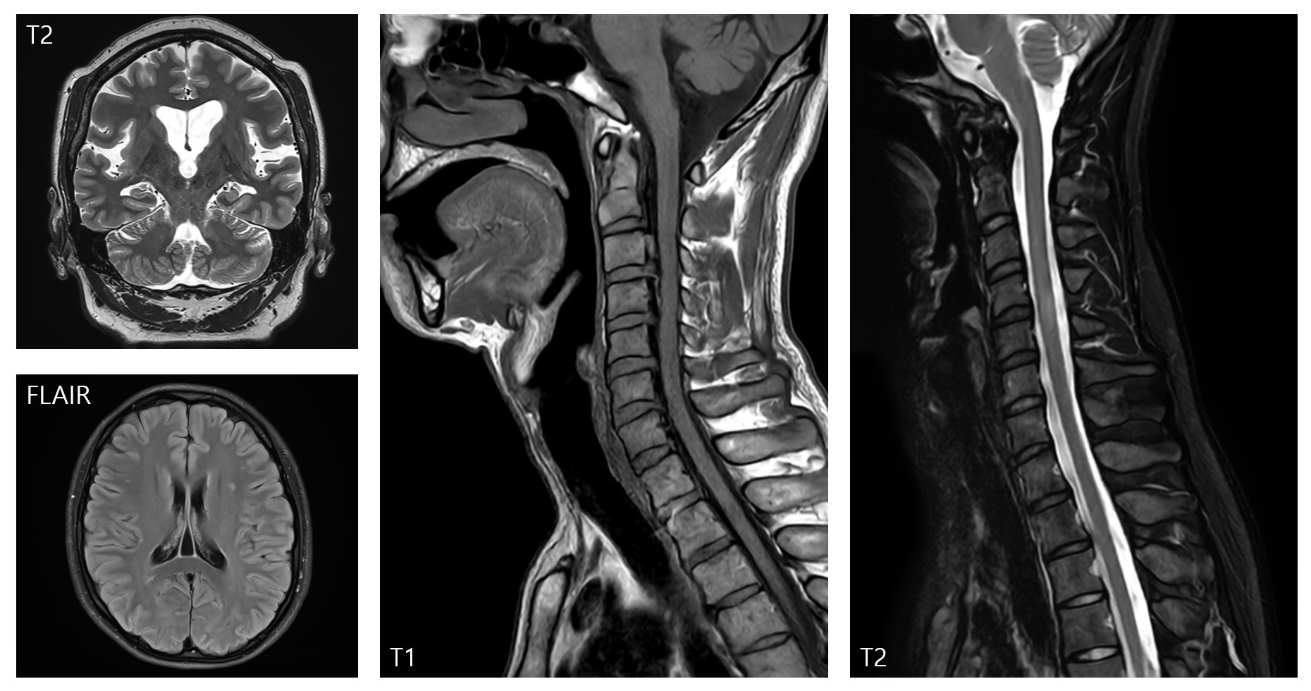
[They all look black and white but not the same…]
At this point, it seems like the filming time is getting longer for a very complicated reason because the A.3-dimensional human body is being filmed in a single test B. from various directions C. with various techniques. It’s worth recalling that all of these stories are about MRI, one of the radiology tests. Even when you take pictures daily, if you don’t set your goals properly, you may have to retake them because they are out of focus or indistinct. MRI is a radiology test used to diagnose our body’s condition and conduct medical activities at medical institutions, so it is very important to take pictures with “medically diagnosable image quality” so that even small lesions are not missed. Therefore, we would like to capture as much information as possible in the test even if it takes a little time to obtain dense and detailed information on the shape and condition of the human structure during the above various filming.
Looking at it so far, there are various reasons for the long MRI examination, so it is strange whether the examination can be completed in about 30-40 minutes, and many medical institutions have standardized the filming method to some extent for various areas and lesions. You decide which direction you will take and how many images you will have, so you don’t have to worry too much about the patient and get the minimum amount of images you need. However, as much detail as it contains, we are still guided not to move during filming as we conduct MRI tests for a long time, which is also difficult for healthy adults.
Through the development of MRI devices, efforts have been made to shorten MRI imaging time through various technologies.
AIRS Medical’s SwiftMR is also a medical device software that enables high-quality images* that can be used in clinical trials based on a new technology called AI deep learning. Why don’t you experience SwiftMR, which reduces existing MRI scan time by up to half, giving patients rapid MRI scans and comfort and increasing examination efficiency for healthcare workers to provide a better medical experience for everyone?
* Approved or registered in 10 countries, including KFDA (class I), U.S. FDA 510k
References: Prince, J.L., Links, J.M., Medical Imaging: Signals and Systems, Pearson Education, 2006